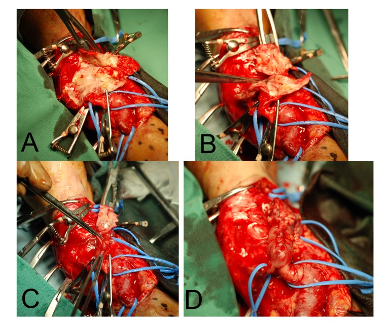
Fig. 3.
The picture of the surgical procedure. A: The proximal and distal portions of the AVF were clamped and the aneurysm was opened. B: The aneurysm was resected, and the back wall was approximated partially with a polypropylene suture. C: A plastic tube was placed inside the AVF and the AVF was closed with polypropylene sutures after the resection of redundant side wall. D: Completion of the remodeling. AVF, autogenous arteriovenous fistula.

