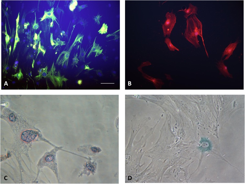
FIGURE 2.

(A) Quiescent PSCs stain positively for desmin by IF. (Hoechst 33258 staining, and Alexa Fluor 488 staining for desmin, original magnification, ×200). (B) Activated PSCs stain positively for α-SMA by IF (Hoechst 33258 staining, and Alexa Fluor 594 staining for α-SMA, original magnification, ×200). (C) Quiescent PSCs have an angular appearance, contained lipid droplets by oil red O staining. (Original magnification, ×400). (D) Senescence is shown in PSCs with cytoplasmic blue staining of SA-β-gal. (Immunocytochemistry; original magnification, ×400) (Xue et al., 2017b).
