FIGURE 6.
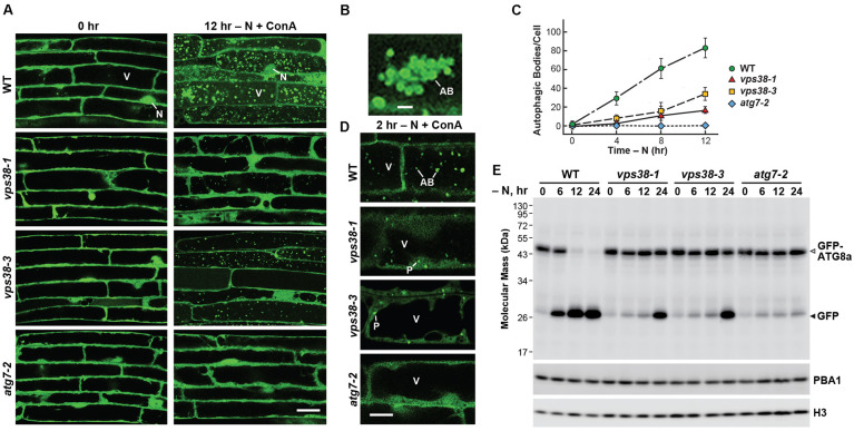
vps38 mutants display defects in autophagy. (A) vps38 mutants accumulate fewer autophagic bodies upon nitrogen starvation. Seven-day-old WT, vps38-1, vps38-3, and atg7-2 seedlings were grown under a LD photoperiod on nitrogen-rich solid MS medium, transferred to nitrogen-deficient liquid MS medium containing 1 μM ConA, and incubated in the light for 12 h. Root cells were imaged by confocal fluorescence microscopy. Scale bar = 20 μm. V, vacuole; N, nucleus. (B) Close-up image of a cluster of autophagic bodies that accumulate in WT vacuoles starved for nitrogen as in (A). Scale bar = 5 μm. (C) Time course for the accumulation of autophagic bodies upon nitrogen starvation. Roots were treated as in (A). The accumulation of autophagic bodies were quantified by confocal microscopy and expressed as a mean (±SD) of at least 25 cells. (D) vps38 mutants accumulate GFP-ATG8a-decorated autophagic structures in the cytoplasm but poorly assemble autophagic bodies. The seedlings were grown as in (A) but incubated in nitrogen-deficient medium containing 1 μM ConA for only 2 h. Scale bar = 5 μm. AB, autophagic body. P, possible phagophore/autophagosome. V, vacuole. (E) vps38 mutants display defects in autophagic transport as judged by the release of free GFP from GFP-ATG8a (Chung et al., 2010). Seedlings were grown under continuous light on nitrogen-rich MS liquid medium, transferred to nitrogen-deficient liquid MS medium, and then harvested at the indicated times. Total seedling extracts were immunoblotted with anti-GFP antibodies, using anti-PBA1 and anti-histone H3 antibodies to confirm near equal protein loading. Open and closed arrowheads locate the GFP-ATG8a fusion and free GFP, respectively.

