Figure 8.
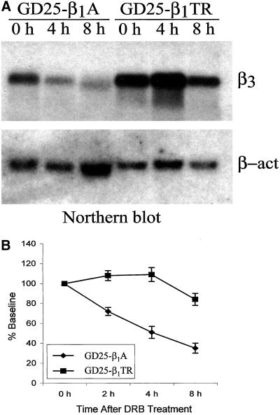
The presence of the β1 cytoplasmic domain doubles the decay rate of the β3 mRNA in GD25 cells. GD25-β1A and GD25-β1TR cells grown to confluence were divided into three 10-cm Petri dishes and cultured in DMEM containing 10% FBS for 24 h before the addition of 60 μM DRB, an inhibitor of transcription initiation. After addition of DRB, the cells were collected at 0, 4, and 8 h for RNA analysis. Total RNA isolation and Northern analysis were performed as described in MATERIALS AND METHODS. (A) Northern blot: equal amounts of total RNA (25 μg/lane) were probed sequentially by 32P-labeled mouse integrin β3 and β-actin cDNA fragments. (B) Scanning densitometry analysis of β3 mRNA levels as detected by Northern blot. β3 Northern signals were normalized to β-actin and displayed as percentage of the baseline (time 0). Data presented are the mean values ± SE of three independent experiments. Notice the lower stability of β3 integrin subunit mRNA in GD25-β1A than in GD25-β1TR cells.

