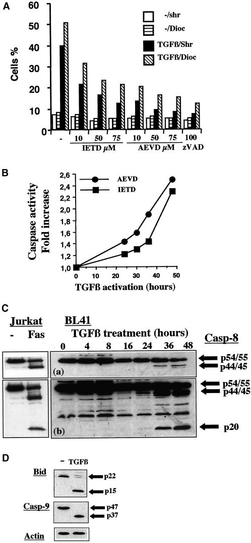
Figure 2.
TGFβ promotes the caspase-8 dependent loss of ΔΨm and apoptosis. (A) BL41 cells were cultured for 48 h without (−) or with TGFβ (1 ng/ml) and various concentrations of IETD-fmk (10, 50, 75 μM), AEVD-fmk (10, 50, 75 μM), or ZVAD-fmk (100 μM). Cell shrinkage (Shr) and the loss ΔΨm (Dioc) were assessed by flow cytometry, as described in Figure 1. (B) Cells were cultured with TGFβ (1 ng/ml) and caspase activity was determined at various times after TGFβ treatment, with the use of IETD-pNA and AEVD-pNA as substrates. Results are expressed as the ratio between caspase activity in TGFβ-treated cells and that in control cells. (C) Cells were cultured with TGFβ (1 ng/ml) for various periods of time. Whole cell extracts were separated by SDS-PAGE and the various forms of caspase-8 were detected by immunoblotting with the use of anticaspase-8 antibodies. A short (a) and a longer (b) exposure of the films is shown. As control for caspase-8 specificity, Jurkat cells were cultured for 24 h in the absence (−) or the presence of anti-Fas antibody (CH11 antibody, 1 μg/ml). (D) Cells were cultured without (−) or with TGFβ (1 ng/ml) for 48 h. Whole cell extracts were separated by SDS-PAGE and the levels of the various forms of Bid and caspase-9 were determined by immunoblotting, with the use of anti-Bid or anti-caspase-9 antibodies. Results are representative of three independent experiments. The amount of protein loaded in each lane was assessed by stripping the filter and reprobing it with an antibody specific for human actin.

