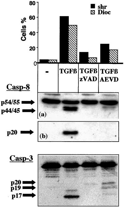
Figure 3.
AEVD inhibits TGFβ-mediated caspase-8 and caspase-3 activation. BL41 cells were cultured for 48 h without (−) or with TGFβ (1 ng/ml) in association with ZVAD-fmk (50 μM), IETD-fmk (75 μM), or AEVD-fmk (75 μM). Cell shrinkage and loss of ΔΨm were assessed by flow cytometry, as described in Figure 1. The cleavage of caspase-8 and of caspase-3 was assessed by immunoblotting. A short (a) and a longer (b) exposure of the films is shown for caspase-8. Results are representative of three independent experiments.

