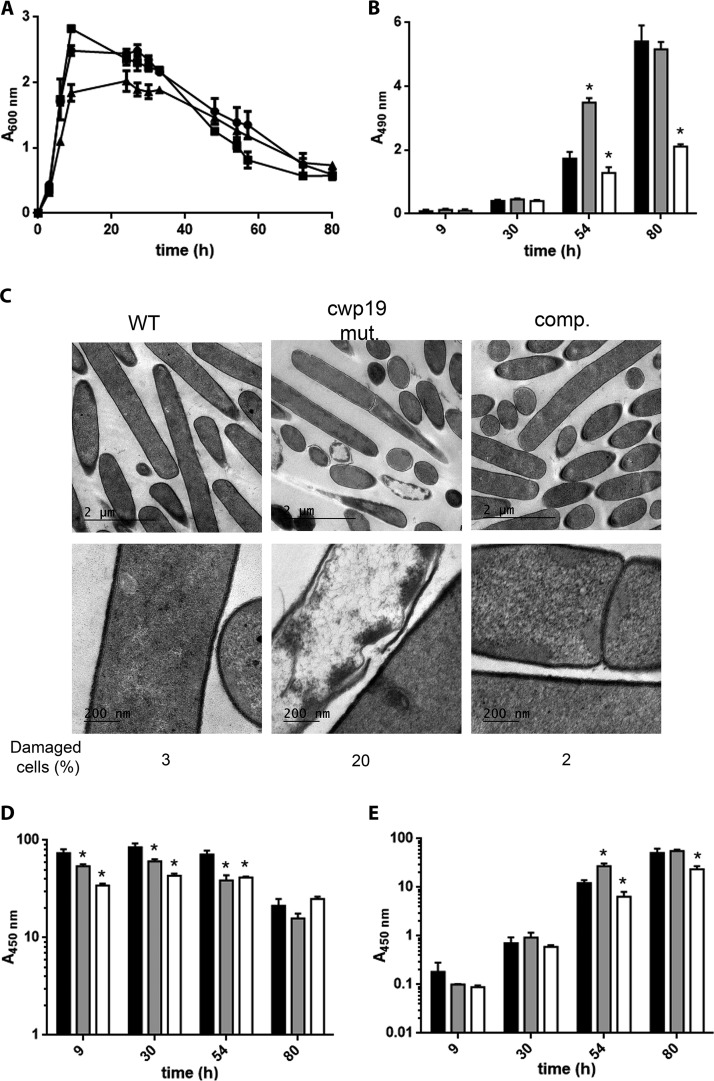
FIG 5 .
Role of Cwp19 in cell autolysis and toxin release of C. difficile cultivated in TY medium. (A) Growth and autolysis of wild-type 630Δerm (●), cwp19 mutant (■), and complemented cwp19 mutant (▲) strains in TY medium. Values are given as means ± standard deviations (n = 3). (B) LDH activity released in the supernatant fractions of wild-type 630Δerm (black bars), cwp19 mutant (gray bars), and complemented cwp19 mutant (white bars) strains in TY medium at different time points. LDH activity was determined using Promega CytoTox 96. The signal from the test was recorded as absorbance at 490 nm. Values are given as means ± standard deviations (n = 3). *, P ≤ 0.05 (Student’s t test). (C) TEM micrographs of C. difficile wild-type 630Δerm, cwp19 mutant (cwp19 mut.), and complemented cwp19 mutant (comp.) strains incubated for 48 h in TY medium. The percentage of damaged cells for each strain is indicated. (D and E) Toxin titers in the cytosol (D) and in the culture supernatant (E) of wild-type 630Δerm (black bars), cwp19 mutant (gray bars), and complemented cwp19 mutant (white bars) strains grown in TY medium. Toxins were quantified by ELISA. The signal from the test was recorded as absorbance at 450 nm. Values are given as means ± standard deviations (n = 3). *, P ≤ 0.05 (Student’s t test).

