FIG 2 .
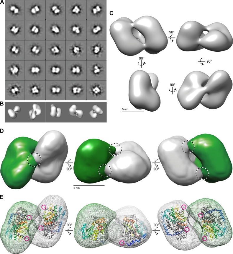
3D structure of the UL69 tetramer determined by 3D reconstruction of negative-stain EM images. (A) A selection of 25 projection-class averages were calculated using reference-free multivariate statistical analysis (MSA). Box size = 33 nm. (B) Surface-rendered views of the final 3D reconstruction of the UL69long envelope orientated to match the bottom row initial class averages of panel A. (C) Surface-rendered views of the UL69 long structure. Five-nanometer scale bars are shown. (D) Segregation of the UL69long structure into two lobe volumes related by C2 symmetry and colored green and gray. Proposed bridge features are highlighted by dashed circles. (E) Each lobe of UL69 was fitted to the ICP27 IHD dimer coordinates. The surface of UL69long is shown as a mesh, and ICP27 (PDB accession number 4YXP) is shown as a ribbon trace, with one chain of the homo-dimer colored gray and the second chain in rainbow colors (blue to red, from the N terminus to the C terminus). The N termini of ICP27 are further highlighted by magenta circles, highlighting their proximity in space to each other and to the bridge regions.

