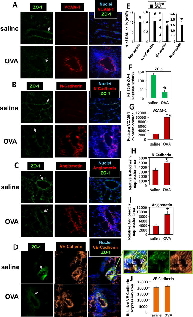
Figure 1. Allergic lung inflammation induces a decrease in ZO-1 in lung endothelial cells and increased lung endothelial cell expression of VCAM-1, N-Cadherin and angiomotin.

Mice were sensitized and challenged with OVA or saline and then lungs were lavaged and lungs in OCT were stored frozen at -80°C. A-D) Lung tissue sections were prepared and labeled with rat anti-mouse VCAM-1, rabbit anti-mouse N-Cadherin, rabbit anti-mouse angiomotin or rat-anti-mouse VE-Cadherin followed by Texas red-conjugated secondary antibodies. Then the sections were labeled with FITC-conjugated anti-mouse ZO-1. Representative wide-field fluorescence micrographs are shown. Panel D has an enlarged image depicting a slice of the vessel in the section that is at a slight oblique angle through the vessel because this nicely encompassed and thus depicted more of the cell junctions. The vascular structure in the lung tissue sections was confirmed by characteristic morphology of vessels in the lung tissue sections (data not shown). Arrow indicates venule. E) Lung lavage cells. F-J) Relative sum of fluorescence pixel intensities per area of the venule endothelial cells. Isotype control antibody-labeled cells were negative (data not shown). N=8-10 mice. Presented as mean ±SEM. *, p<0.05.
