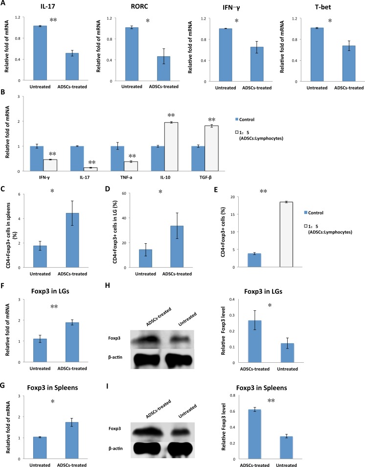
Figure 5.
Adipose-derived MSC treatment regulated Th1/Th17/Tregs in vivo and in vitro. (A) Lacrimal glands were removed from untreated and ADSC-treated rabbits, and IL-17, IFN-γ, T-bet, and RORC mRNA expression was analyzed by real-time qPCR. (B) Adipose-derived MSCs cocultured with splenic lymphocytes at the ratio of 1:5 were collected. The mRNA expression levels of IL-10, IFN-γ, IL-17, TNF-α, and TGF-β were measured by qPCR. (C) Percentage of CD4+Foxp3+ cells among splenic lymphocytes isolated from ADSC-treated and untreated rabbit spleen. (D) Percentage of CD4+Foxp3+ cells among lymphocytes isolated from LGs of ADSC-treated and untreated rabbits. (E) Adipose-derived MSCs cocultured with splenic lymphocytes at the ratio of 1:5 were measured by flow cytometry for the percentages of CD4+Foxp3+ cells at day 5. (F, G) Quantitative PCR analysis of Foxp3 mRNA expression in LGs and spleens of ADSC-treated and untreated rabbits. (H, I) Immunoblot of LGs and spleens of ADSC-treated and untreated rabbits. Tissue lysates were immunoblotted with indicated antibodies. Left: Levels of Foxp3 and β-actin. Right: Relative Foxp3 level = Foxp3 level/β-actin level. Data are representative of three independent experiments and bar graphs show mean ± SD, n = 3. *P < 0.05, **P < 0.01.

