Figure 4.
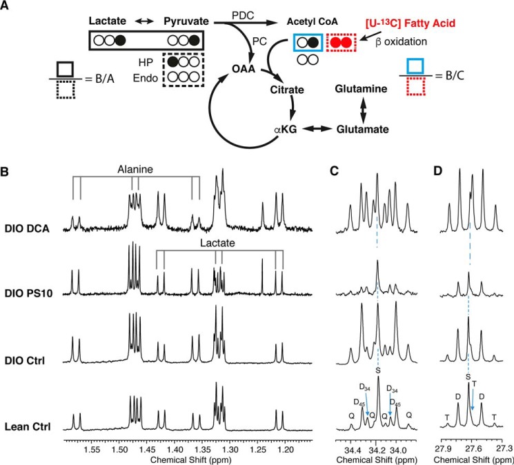
The proton NMR spectrum of alanine, lactate, and 13C labeling patterns of glutamate from the mouse hearts with different PDK inhibitor treatments. A, sources of 13C label on acetyl-CoA and glutamate in the TCA cycle. The ratio of labeled pyruvate/lactate over unlabeled endogenous sources is defined as B/A. The ratio of labeled pyruvate/lactate over labeled fatty acid is defined as B/C. B, the proton spectrum of methyl group on alanine and lactate. The main doublet (center two lines, 12C bonded 1H resonances) and 13C directly bonded satellite doublets (on two ends) are indicated. The 4JCH manifests as doublets close to the 12C-bonded resonances around 1.33 ppm and 1.47 ppm. C and D, the 13C signals of label enrichment on glutamate C4 (C) and glutamate C3 (D) are shown. The coupling pattern is shown as S, singlet; D, doublet; T, triplet; Q, quartet. The subscript numbers indicate carbons of the coupling. HP, hyperpolarized [1-13C]pyruvate. Endo, unlabeled pyruvate derived from endogenous sources. Note that no assumption of steady state is needed for this analysis.

