
Figure 6.
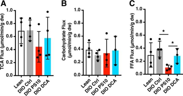
PS10 and DCA did not alter TCA flux in DIO mouse hearts as measured with a non–steady state isotopomer analysis. A, TCA flux of hearts with different treatment. B, the absolute carbohydrate contribution to TCA cycle flux in the mouse hearts with different treatment. C, the absolute free fatty acid contribution to TCA cycle flux in the mouse hearts with different treatment. Data are presented as mean ± S.D. n = 4 all groups. *, p < 0.05.