Figure 2.
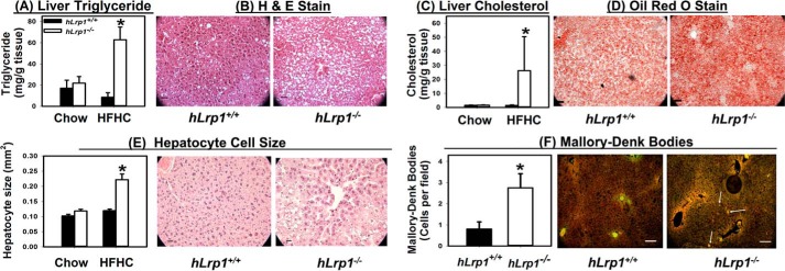
Hepatic LRP1 deficiency promotes steatosis, hepatomegaly, and Mallory–Denk body accumulation. Livers were excised from hLrp1+/+ (filled bars) and hLrp1−/− (open bars) mice after feeding a low-fat, low-cholesterol chow or the HFHC diet for 16 weeks. Hepatic steatosis was assessed after lipid extraction and quantification of triglycerides (A) and cholesterol (C) from eight liver samples from each group. Liver sections from HFHC-fed mice were also processed for histological staining. Representative images of hematoxylin and eosin (H&E)-stained sections showed the accumulation of lipid vacuoles (B), and Oil Red O–stained sections showed lipid accumulation (D) (right panels). E, the stained liver sections also revealed an increase in hepatocyte cell size (30–40 cells measured in each field). F, the liver sections were also used for immunofluorescence detection of keratin 8/18. Representative images are shown in the right panel, with white highlighting of selected cells with Mallory–Denk bodies. The number of cells with Mallory–Denk bodies were counted (from 16–20 mice in each group) and reported as a bar graph in the left panel. *, significant difference from hLrp1+/+ mice at p < 0.01. Scale bar in each image, 50 μm.

