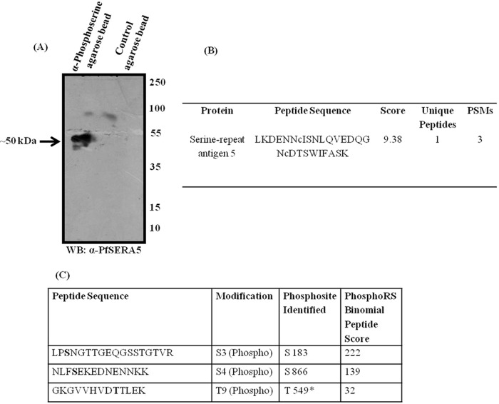
Figure 1.
PfSERA5 is phosphorylated at the schizont stage in P. falciparum. A, representative immunoblot showing a band of ∼50 kDa in eluates of the affinity pulldown from P. falciparum schizont extracts using anti-phosphoserine antibody when probed with anti-PfSERA5 mouse serum. WB, Western blot. B, identification of PfSERA5 from the LC-MS/MS analysis of the above mentioned immunoprecipitates. Other proteins identified by this pulldown are summarized in Table S1. PSMs, peptide spectrum matches. C, the sites of phosphorylation in PfSERA5 as identified by LC-MS/MS analysis after immunoprecipitation using anti-PfSERA5 serum from P. falciparum schizont extracts.

