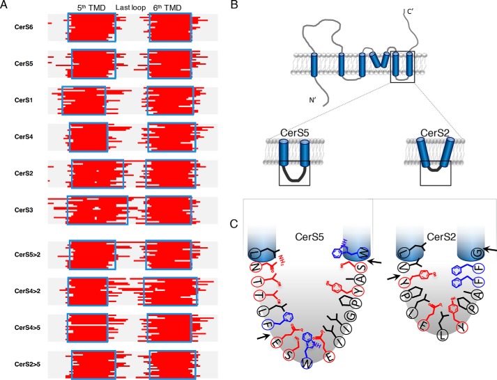
Figure 7.
Residues in the loop between TMD5 and -6 in CerS2 and CerS5. A, predictions of the fifth and sixth TMDs (red) by 19 different programs in the six human CerS (top) and chimeric proteins (bottom). Blue rectangles indicate residues where the majority of the programs (>10/19) predict a TMD. See also Fig. S7A. B, conformation of TMDs 5 and 6 might be restricted according to the loop length. C, residues of CerS5 (left) and CerS2 (right) within the last putative loop. The arrows indicate the region that was altered between CerS5 and CerS2. Polar residues are indicated in red and bulky residues in blue.

