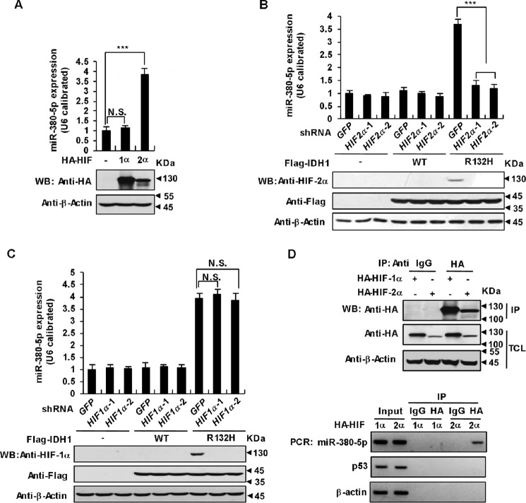
Figure 6.
HIF-2α promotes miR-380-5p transcription. A, overexpression of HIF-2α up-regulates miR-380-5p. HCT116 cells were transfected with HA-tagged HIF-1α or HIF-2α. 36 h after transfection the expression of miR-380-5p was analyzed with qRT-PCR and normalized to U6 (mean ± S.D., n = 3, ***, p < 0.001, unpaired Student's t test). B, knockdown of HIF-2α down-regulates miR-380-5p. HCT116 cells stably expressing WT IDH1 or its R132H mutant were infected with lentiviruses expressing two independent HIF-2α shRNAs or control shRNA, individually. 48 h after infection, the expression of miR-380-5p was analyzed and presented as in A (***, p < 0.001). C, knockdown of HIF-1α has no effect on miR-380-5p up-regulation caused by the IDH1 Arg-132 mutant. HCT116 cells stably expressing WT IDH1 or its R132H mutant were infected with lentiviruses expressing two independent HIF-1α shRNAs or control shRNA, individually. 48 h after infection, the expression of miR-380-5p was analyzed and presented as in A (N.S. = not significant). D, HIF-2α binds to the promoter regions of miR-380-5p. HCT116 cells were transfected with HA-tagged HIF-1α or HIF-2α. 36 h after transfection, ChIP assays were performed using IgG (control) or anti-HA antibodies. Proteins in precipitates and total cell lysates were determined by Western blot analysis (upper panel). Purified DNA was analyzed by standard PCR using primers targeting the indicated promoter regions (lower panel). Actin and p53 were used as negative controls.

