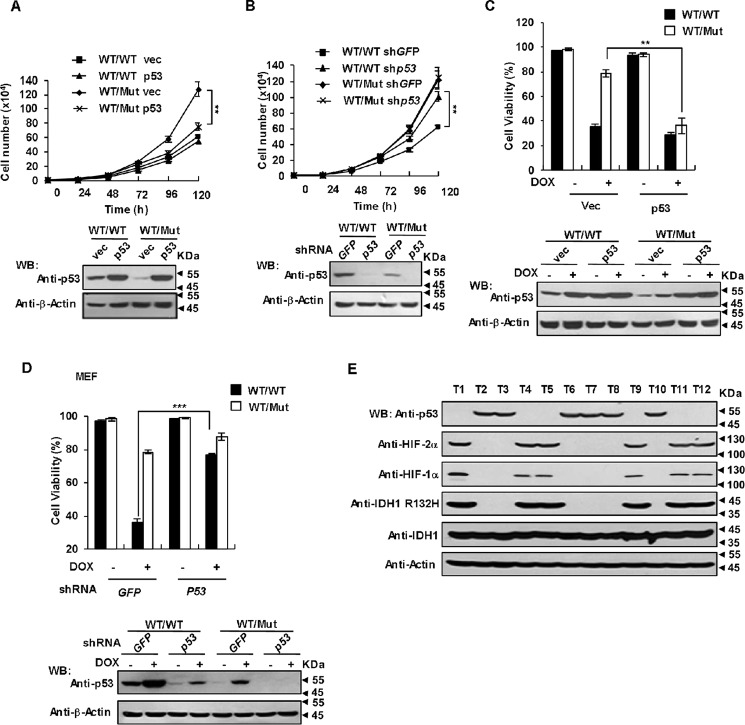
Figure 7.
p53 down-regulation is involved in IDH1 mutant-driven tumorigenesis. A, rescue expression of p53 significantly attenuated the proliferation rate of IDH1 Arg-132 mutant cells. Both IDH1WT/WT and IDH1WT/Mut cells were infected with lentiviruses expressing p53. At 36 h after infection, proliferation rates were determined by growth curves (upper panel). Mean ± S.D., n = 3 independent experiments, are shown (**p < 0.01, unpaired Student's t test). Proteins in total cell lysates of the same cell lines were determined by Western blot analysis (WB) (lower panel). B, knockdown p53 increased the proliferation rate of IDH1WT/WT cells but not IDH1WT/Mut cells. IDH1WT/WT and IDH1WT/Mut MEFs were infected with lentiviruses expressing p53 shRNA or control shRNA. At 36 h after infection, proliferation rates (upper panel) were determined as in A (**, p < 0.01, unpaired Student's t test). Proteins in total cell lysates of the same cell lines were determined by Western blot analysis (lower panel). C, forced expression of p53 sensitize IDH1 Arg-132 mutant cells to DOX-induced apoptosis. IDH1WT/WT and IDH1WT/Mut MEF cells were infected with lentiviruses expressing p53. At 36 h after infection, cells were treated with or without 2.5 μm DOX for 16 h. The percentages of surviving cells (Annexin V negative) were determined by a flow cytometer. Data are presented as mean ± S.D. of three independent experiments (**, p < 0.01, unpaired Student's t test). D, knockdown of p53 desensitized of IDH1WT/WT cells rather than IDH1WT/Mut cells to DOX-induced apoptosis. IDH1WT/WT and IDH1WT/Mut MEF cells were infected with lentiviruses expressing p53 shRNA. At 36 h after infection, cells were treated with or without 2.5 μm DOX for 16 h. The percentages of surviving cells were determined and presented as in D (***, p < 0.001, unpaired Student's t test). E, IDH1 R132H mutant suppresses p53 expression in glioma. Clinical specimens of glioma were analyzed by Western blot with the antibodies as indicated.

