Figure 2.

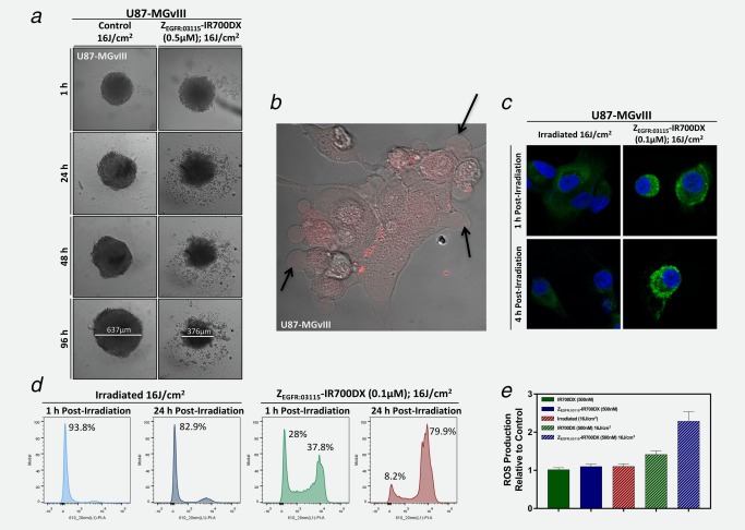
In vitro morphological changes following affibody‐based PIT. (a) Incubation of U87‐MGvIII spheroids with the ZEGFR:03115–IR700DX for 6 h and irradiation with a red LED (16 J/cm2) induced phototoxic cell death and disintegration of the architectural structure of the spheroid population. (b) U87‐MGvIII cells grown as a monolayer culture showed rapid cell swelling and bleb formation (see arrows) as visualized by a phase‐contrast image 1 h post ZEGFR:03115–IR700DX (red) irradiation with the 639 nm laser on a confocal microscope. (c) Following methanol fixation of U87‐MGvIII cells, either treated by PIT or just irradiated, and staining with an anti‐calreticulin‐AlexaFluor488 antibody overnight (4°C), images were acquired by confocal microscopy. (d) Cell membrane disruption was monitored by propidium iodide (1 µg/mL) staining. U87‐MGvIII cells irradiated only or treated with ZEGFR:03115–IR700DX‐based PIT were analyzed by flow cytometry 1 and 24 h post‐treatment. (e) Reactive oxygen species production was assessed using the DCFDA cellular ROS detection assay kit using U87‐MGvIII cells treated with affibody‐based PIT (15 min after light exposure). The results were normalized to the control cells. Data are presented as mean ± SEM (n = 3).
