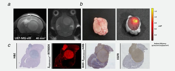
Figure 5.

ZEGFR:03115–IR700DX accumulates in U87‐MGvIII orthotopic glioma tumors. (a) T2‐weighted MRI images of an intracranial brain tumor model 11 days post‐cell implantation. (b) Photographic image of the brain and the corresponding ZEGFR:03115–IR700DX fluorescent image demonstrates predominant accumulation of the conjugate within the brain tumor mass. (c) Transaxial brain histological sections (10μm) containing tumor tissue were obtained for ex vivo analysis immediately after 1 h in vivo image acquisition. ZEGFR:03115–IR700DX clearly delineated tumor mass from the surrounding normal tissues which correlated well with H&E and EGFR staining of the consecutive sections.
