
Figure 1.
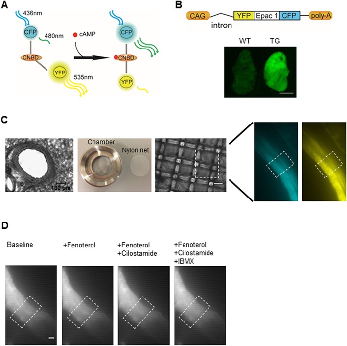
Monitoring cAMP FRET signals using lung slices of CAG‐Epac1‐camps transgenic mice. (A) An Epac1‐based FRET sensor for real‐time cAMP detection. This cAMP biosensor consists of CPF and YFP flanking a cyclic nucleotide binding domain (CNBD) from Epac1. Binding of cAMP leads to a conformational change, allowing an increase of CFP and a decrease of YFP fluorescence to be observed. (B) CAG‐Epac1‐camps transgenic mice express Epac1‐camps sensor ubiquitously, under the control of the CAG promoter. Fluorescent image of the superior lung lobe of a transgenic mouse compared with that of a wild type counterpart. Scale bar: 3 mm. (C) Slices of 200 μm thickness were cut using a vibratome. A lung slice was placed in the custom‐made image chamber, and 600 μL of FRET buffer was added into the chamber, and a home‐made net was used to fix the slice in the chamber in order to reduce airway movement artefacts. The fluorescent images (480 nm: left, 535 nm: right) of intrapulmonary airway are observed under inverted fluorescent microscope (×40). The region of interest is indicated in the white boxes. (D) Representative images showed no significant airway movement after treatment with fenoterol alone or in combination with cilostamide and IBMX. Similar images were observed with rolipram (not shown). Scale bar: 20 μm.