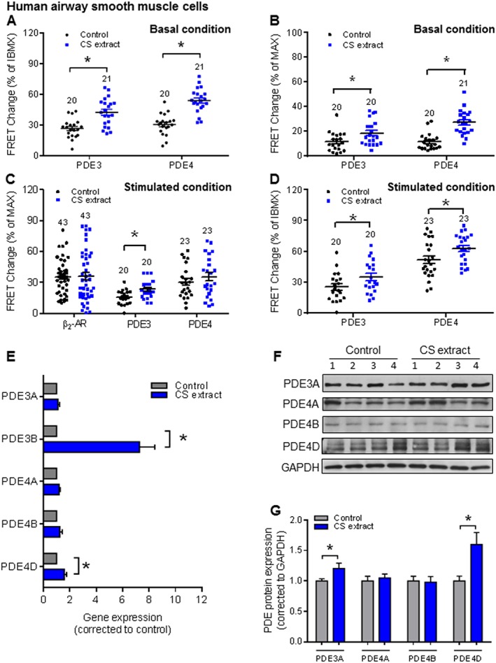
Figure 5.

CS extract induces PDE3 and PDE4 changes in HASM. HASM cells were incubated with control medium or medium containing CS extract (2.5% v/v; final concentration) for 24 h. FRET data analysis of HASM (A–D) under basal conditions (A, B) and fenoterol stimulated conditions (C, D). Quantification of basal PDE inhibitor responses revealed that both PDE3 and PDE4 were significantly up‐regulated by CS extract (A, B). We observed a significant increase of PDE3 contributions induced by CS extract after β2‐adrenoceptor stimulation, while fenoterol‐induced and rolipram‐induced FRET responses were not affected (C). In HASM cells, CS extract significantly increased both PDE3 and PDE4 profiles in total PDEs (D). Number of independent experiments is indicated above the bars. (E) Gene expression of PDE3 (PDE3A and PDE3B) and PDE4 subfamilies (PDE4A, PDE4B and PDE4D) was analysed by real‐time quantitative PCR in lung slice lysates. CS extract increased the gene expressions of both PDE3B and PDE4D. (F, G) Protein expression of PDE3A and PDE4 subfamilies (PDE4A, PDE4B and PDE4D) were determined by Western blot analysis in lung homogenates. Quantification and representative blots are shown. Data are from nine independent experiments. Data are expressed as mean ± SEM, *P < 0.05; significantly different from control; One‐way ANOVA was used for simple two groups comparison. Rolipram, 10 μM; cilostamide, 10 μM; IBMX, 100 μM; fenoterol, 100 nM for HASM cells.
