Figure 3.
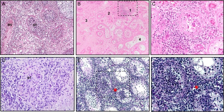
Histopathology of human orchitis of different etiology and mouse experimental autoimmune orchitis. (A) Human testis: acute bacterial orchitis (epididymo-orchitis) with massive infiltration of both the interstitium and seminiferous tubules (ST) with inflammatory cells, including numerous neutrophils. The architecture of affected ST is largely disrupted, whereas adjacent ST show hypospermatogenesis; interstitial edema and enlarged venous blood vessel (BV) (Periodic acid–Schiff stain, objective ×10). (B) Sequelae of mumps orchitis with persistent focal inflammation in human testis: Dense peritubular lymphocytic infiltrate involving the lamina propria as well as adjacent blood vessels (1), tubular atrophy resulting in complete hyalinization (‘tubular shadows’; 2, 3), and interstitial fibrosis (3). The adjacent seminiferous tubules show hypospermatogenesis; note the ‘flattened’ epithelium with a complete loss of the adluminal compartment in some tubules (4); (hematoxylin–eosin staining, objective ×10). (C) Higher magnification of area 1 in (B); note the characteristic meshwork pattern of the affected lamina propria; the germinal epithelium is largely disrupted, with only a few germ cells remaining (hematoxylin–eosin stain, objective ×40). (D) Human testis: subacute granulomatous orchitis with residual structures of ST containing inflammatory cells (hematoxylin and eosin stain, objective ×40). (E) Characteristic histopathology of mouse experimental autoimmune orchitis (EAO) showing destruction of testicular morphology with reduced size of ST, loss of germ cells and presence of dense peritubular and interstitial inflammatory infiltrates (marked by asterisk; hematoxylin stain, objective ×20). (F) Mouse EAO, higher magnification (hematoxylin stain, objective ×40) of selected area in (E). (A–D) From Schuppe and Bergmann (2013); reprinted with permission of Springer Nature (License number: 4282971349118).

