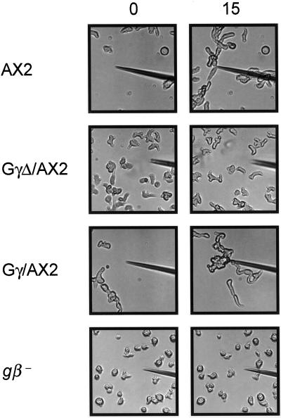
Figure 5.
GγΔ/AX2 cells fail to carry out chemotaxis. Cells at the 6-h stage of development were plated on a cover slide and a micropipette containing 1 μM cAMP was touched to its surface. The cell-movement images were taken by a CCD camera with Zeiss microscope. The left panels were taken when the micropipette first contacted the cover slide. The right panels were taken in 10 min later. Each chemotaxis assay was repeated at least three times.

