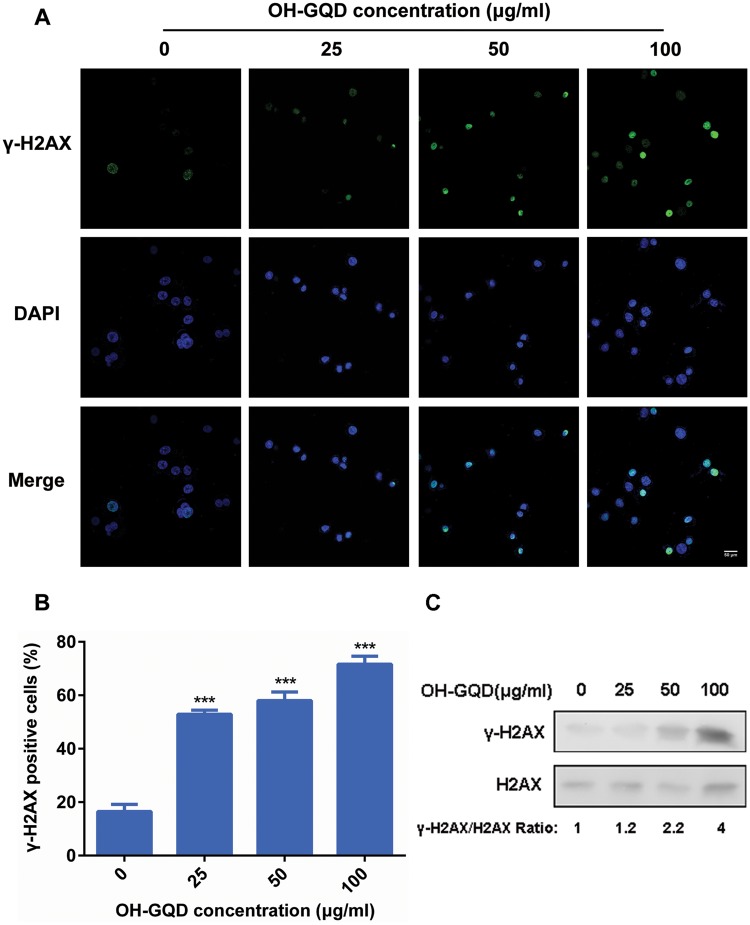
Figure 3.
OH-GQDs induced DNA damage in human esophageal epithelial cells. A, HET-1A cells were treated with the indicated concentration (0, 25, 50, and 100 μg/ml) of OH-GQDs for 24 h; samples were collected and immuno-stained for anti-γ-H2AX antibody. The incidences of γ-H2AX-positive cells were counted. Scale bar = 50 μm. B, Quantitative data are represented as mean ± SD from 3 tests. One-way ANOVA was performed. When compared with 0 μg/ml OH-GQDs-treated cells, *** p < .001. C, Western blot analysis for γ-H2AX and H2AX expressions between OH-GQDs treated and control cells. (For interpretation of the references to colour in this figure legend, the reader is referred to the web version of this article.)

