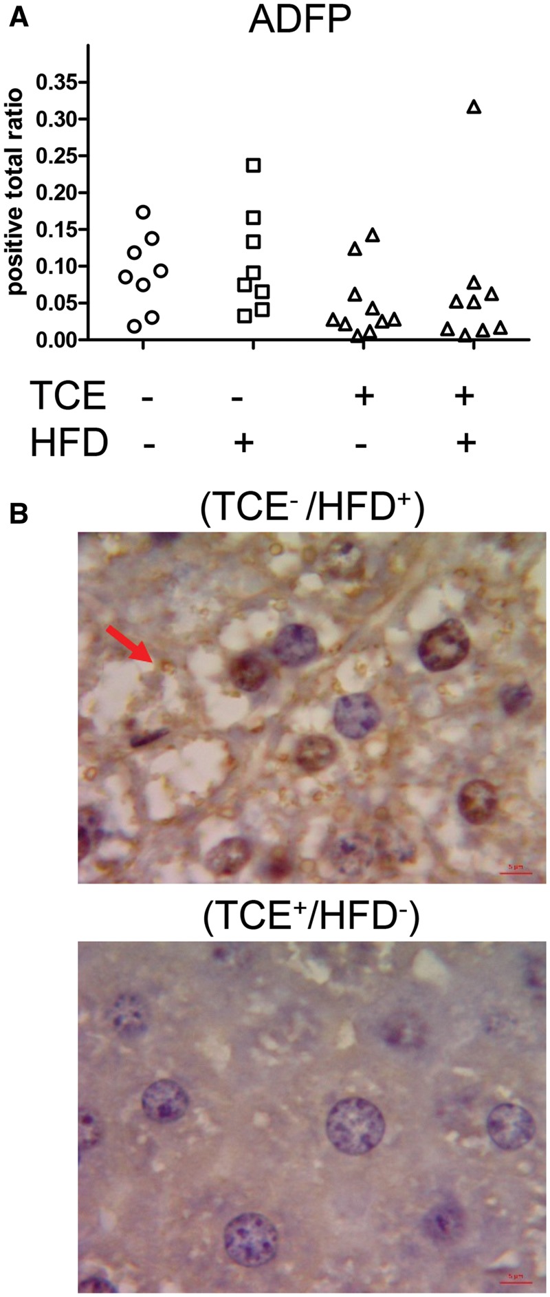
Figure 4.

TCE decreased ADFP staining. Liver tissues were collected from female offspring at study terminus and subjected to Immunohistochemical staining of ADFP or Perilipin-2 as described in the “Materials and Methods” section, quantified using Aperio Positive Pixel Count Algorithm (Leica Biosystems) and represented as positive total ratio. A, Each plot in the graph represents a liver sample from a single mouse within each group. B, Representative staining (100× oil immersion). The arrow points to the ADFP-stained fat droplets in the HFD-exposure group which are significantly decreased in the TCE group.
