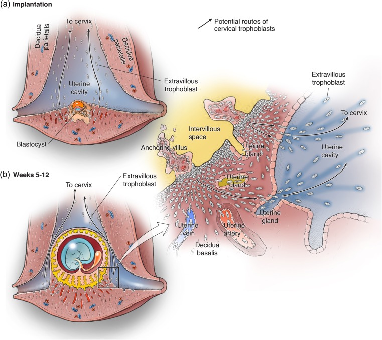
Figure 4.
Origin of cervical trophoblast cells during placental development. The developing conceptus is shown within the uterus at the implantation site (a) and later during the placentation period of weeks 5–12 of gestation (b). EVT cells originate from trophoblast cell columns at the base of the anchoring villi, and follow the interstitial, endovascular, and endoglandular invasion routes. The inset in (b) is expanded to the right, showing the transitional zone of the decidua basalis at the margin of the placenta. As demonstrated in Figs 1 and 2, interstitial EVT cells can invade and replace the uterine epithelium from the basal side, and then enter the uterine cavity. In the decidua basalis, endoglandular EVT cells invade and reach the lumen of UGs. We speculate that, at the lateral margin of the placenta, they are transported together with the glandular secretions into the uterine cavity. Once EVT cells have reached the uterine cavity, they could migrate towards the cervix, or be carried there by the uterine secretion products (arrows).

