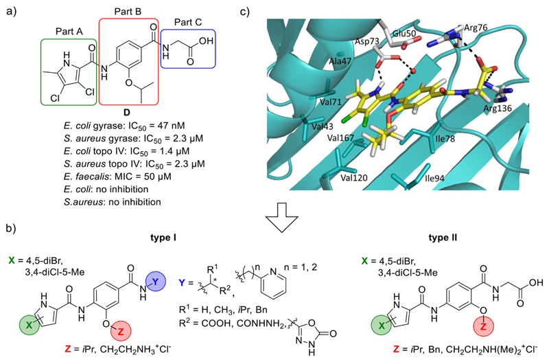
Fig. 2.
a) A representative N-phenylpyrrolamide D and its inhibitory activities on DNA gyrase and topoisomerase IV [16]; b) Structures of type I and type II N-phenylpyrrolamide inhibitors; c) Docking binding mode of inhibitor D coloured according to the atom chemical type (C, yellow; N, blue; O, red; Cl, green) in the ATP binding site of E. coli GyrB (in cyan, PDB code: 4DUH [22]). The water molecule is presented as a red sphere. (For interpretation of the references to colour in this figure legend, the reader is referred to the Web version of this article.)

