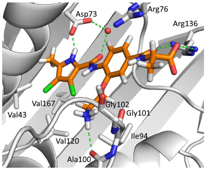
Fig. 3.
The GOLD-predicted binding pose of inhibitor 9e (in orange sticks) in the E. coli GyrB ATP-binding site (PDB entry: 4DUH [22], in grey). Hydrogen bonds are presented as green dashed lines. The water molecule is shown as a red sphere. The figure was prepared with PyMOL [24]. (For interpretation of the references to colour in this figure legend, the reader is referred to the Web version of this article.)

