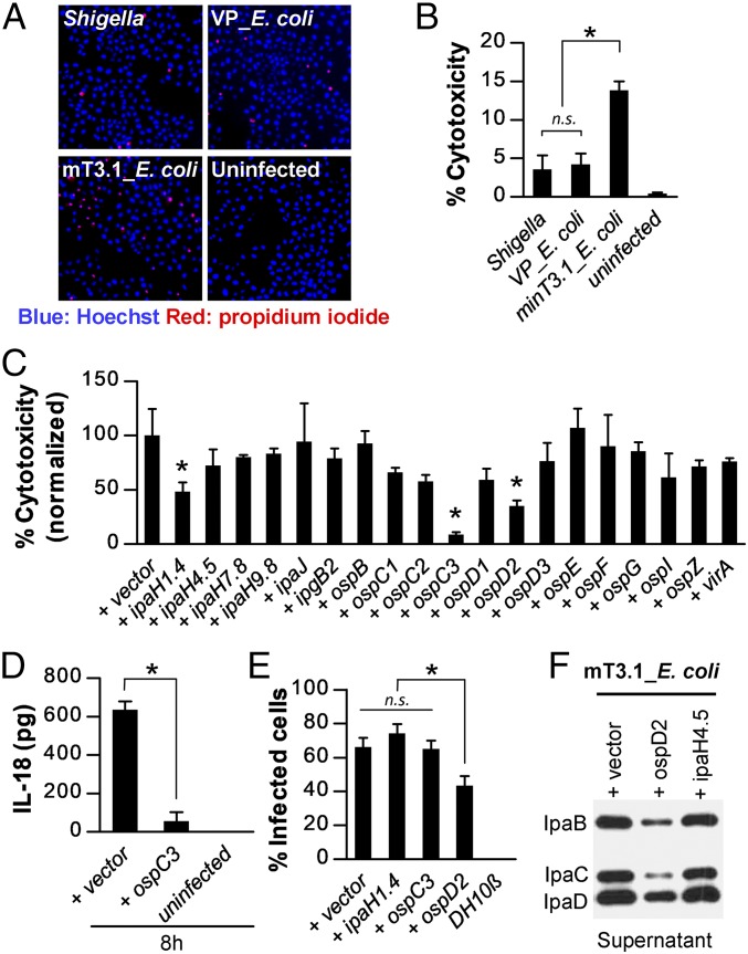
Fig. 2.
mT3.1_E. coli induces epithelial cell death via activation of noncanonical inflammasomes that are suppressed. (A and B) HeLa cells were infected with each strain at an MOI of 100 for 30 min, followed by the addition of gentamicin. After another 2 h, cells were stained with Hoechst (blue) and PI (red). (A) Representative images of stained cells obtained with 4× objective. (B) Quantification of percentage of PI-positive cells (percent cytotoxicity). (C) HeLa cells were infected with mT3.1_E. coli that express each designated effector at an MOI of 100 for 60 min, followed by the addition of gentamicin. After another 1.5 h, the cells were stained with Hoechst and PI. The percentage cytotoxicity normalized to vector control is shown. For each infection, at least 2,000 cells were imaged. *P < 0.05 compared with (+) vector control. (D) Quantification of IL-18 secreted by infected HeLa cells at 8 h postinvasion. (E) At 1 hpi of HeLa cells with each strain at an MOI of 100, the percentage of cells containing intracellular bacteria was determined by an inside/outside microscopy assay, as described in Fig. 1. Data are expressed as the mean ± SD of three experimental repeats. (F) Secretion assays of designated strains conducted as described in Fig. 1. Membranes were immunoblotted with designated antibodies. The blot shown is representative of three experimental repeats. All other data are expressed as the mean ± SD of at least two experimental repeats, each with two technical replicates (n ≥ 4). *P < 0.05 as indicated. n.s., nonsignificant.

