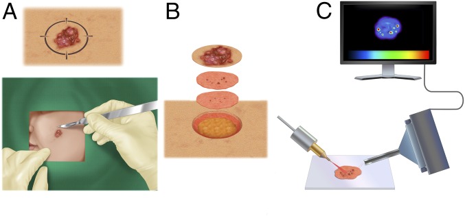
Fig. 1.
General workflow of this study. (A) During Mohs surgery, a surgeon removes a lesion suspected as basal cell carcinoma (BCC). A 2D map of the lesion is created. (B) Using tissue-sparing technique, the visible tumor and a conservative margin of normal skin are removed and subsequently stained for histopathological evaluation. The process continues until the stained sections are clear of microscopic tumor aggregates. (C) Excised skin sections are imaged by desorption electrospray ionization mass spectrometry (DESI-MS) imaging, which can accurately delineate microscopic tumor aggregates without histological staining and evaluation.

