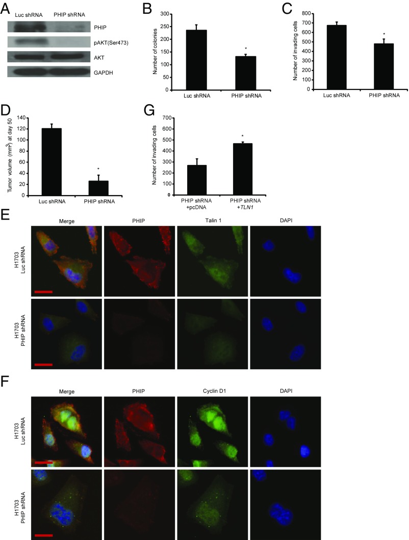
Fig. 3.
Effects of stable shRNA-mediated suppression of PHIP in H1703 cells. (A) Western analysis of expression of PHIP and other proteins in H1703 cells expressing anti-luc shRNA or anti-PHIP shRNA (127738). (B) Colony formation ability of H1703 cells expressing anti-luc shRNA or anti-PHIP shRNA (127738) (P < 0.02). (C) Invasion into Matrigel of H1703 cells expressing anti-luc shRNA or anti-PHIP shRNA (127738) (P < 0.04). (D) Tumor volume 50 d after s.c. injection of H1703 cells expressing anti-luc shRNA or anti-PHIP shRNA (127738) (P = 0.000006). (E and F) Qualitative immunofluorescence analysis of (E) PHIP and Talin 1 and (F) PHIP and Cyclin D1 in H1703 cells expressing anti-luc shRNA or anti-PHIP shRNA (127738). (Scale bar, 20 µm.) (G) Effects of overexpression of TLN1 cDNA or control plasmid on invasive capacity into Matrigel of H1703 cells expressing anti-PHIP shRNA (127738) (P < 0.001).

