Fig. 3.
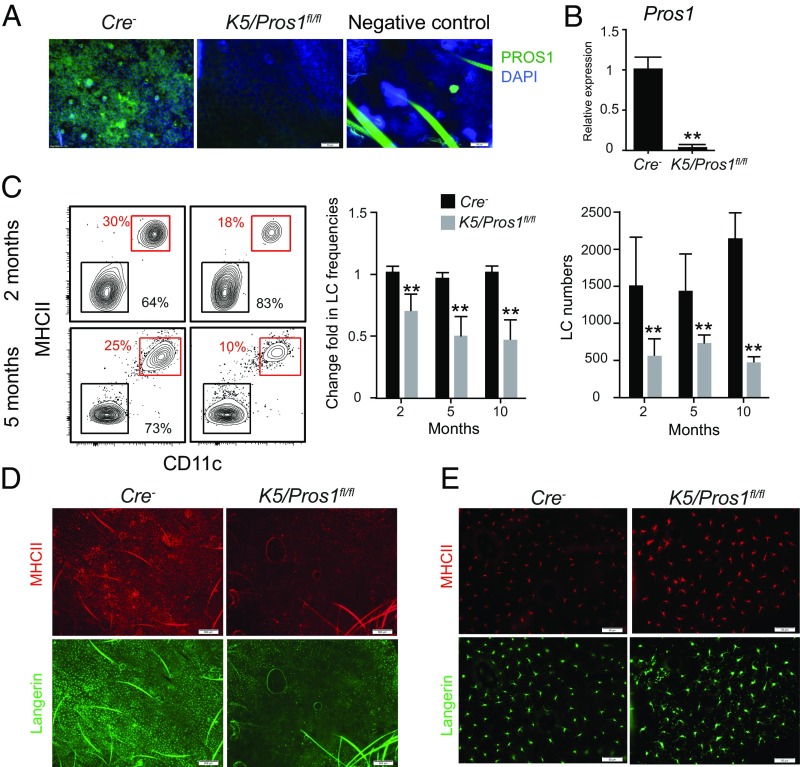
Ablation of PROS1 in keratinocytes reduces LC frequencies. (A) Whole-mount immunofluorescence staining on epidermal layers prepared from adult K5/Pros1fl/fl and littermate Cre− mice, stained for PROS1 (green) and DAPI (blue). Representative fluorescent images are shown, representing one of two independent experiments (n = 3 mice in each experiment). Negative control, primary antibody was omitted. (B) Quantification of mRNA levels of PROS1 in epidermal layers of adult K5/Pros1fl/fl and Cre− mice using RT-qPCR (n = 5 mice). Data of one of two independent experiments are presented as the mean values ± SD. (C) Frequencies and absolute numbers (per a half ear) of LCs (CD45+MHCII+CD11c+ cells) in epidermal cells prepared from 2-, 5-, and 10-mo-old K5/Pros1fl/fl and Cre− mice. Representative flow cytometry plots, as well as bar graphs, present the fold change in LC frequencies (normalized to Cre− mice) or absolute LC numbers in the epidermis of these mice. Data are representative of four independent experiments, and each experiment included at least five separately analyzed mice. (D and E) Whole-mount immunofluorescence staining on epidermal layers prepared from adult K5/Pros1fl/fl and littermate Cre− mice, stained for langerin (green) and MHCII (red). Representative fluorescent images are presented representing one of three independent experiments (n = 3 mice in each experiment). **P < 0.01. (Scale bars: A and E, 50 μm; D, 500 μm.)

