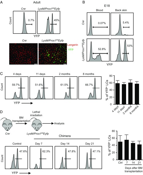
Fig. 7.
Normal development and homeostasis of LCs in LysM/Pros1fl/flEyfp mice. (A) Epidermal cells were prepared from adult LysM/Pros1fl/flEyfp and littermate Cre− mice. Representative flow cytometry plots present the percentages of YFP+ LCs among total epidermal LCs. Representative whole-mount immunofluorescence staining on epidermal sheet stained against langerin (red) and YFP (green). Data are representative of four independent experiments, and each experiment included at least three separately analyzed mice. (B) Blood and back skin epidermis of E18 embryos were sampled from LysM/Pros1fl/flEyfp and Cre− mice. Representative flow cytometry plots present the percentages of YFP+ cells among LC precursors in the blood (CD45+MHCII+) and epidermis (CD45+MHCII+F4/80+). Results of one of two independent experiments are presented. (C) YFP+ LCs were analyzed in the epidermis of LysM/Pros1fl/flEyfp mice at the indicated time points. Representative flow cytometry plots and graph present the frequencies of YFP+ LCs among total LCs (n = 3–5). Data of one of three independent experiments are provided and present the mean values ± SD. (D) Adult LysM/Pros1fl/flEyfp mice were lethally irradiated and, 24 h later, were administered i.v. with BM cells purified from littermate Cre− mice. The epidermis of the chimeric mice was analyzed 7, 14, and 21 d after BM transplantation to quantify the frequencies of YFP+ LCs. Representative flow cytometry plots and graph present the frequencies of YFP+ LCs among total LCs (n = 3). Data of one of two independent experiments are provided and present the mean values ± SD. Ctrl, control. (Scale bars:, 50 μm.)

