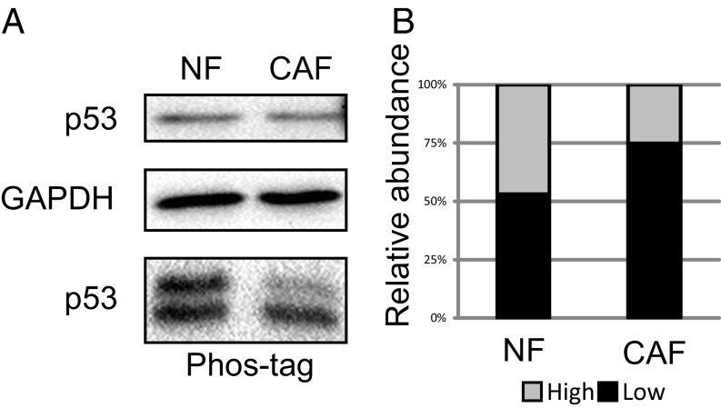
Fig. 2.
CAF p53 is hypophosphorylated. (A) Extracts from immortalized NFs and CAFs (patient 4731) were subjected to either standard SDS/PAGE (Top) or 30 µM Phos-tag SDS/PAGE (Bottom), followed by Western blot analysis with the indicated antibodies. (B) The relative abundance of each band in the Phos-tag gel, denoted by its position in the autoradiogram.

