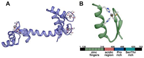
Figure 1. Structures of CaM and MTF1.
(A) Crystal structure of CaM with coordinating ligands highlighted (PDB entry 4BW8). (B) Crystal structure of ZIF-268 as an example of a αββ Zn2+ finger fold. MTF-1 encodes six similar Zn2+ fingers and three transactivation domains as shown in the schematic below the structure (PDB entry 1ZAA).

