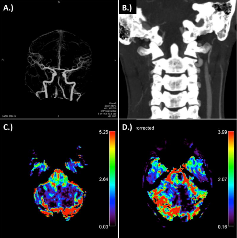
Figure 1.
CT Stroke Protocol at time of presentation. A.) 4D CTA demonstrating top of the basilar occusion and poor flow in the right vertebral artery; B.) coronal CA demonstrating dissection of verterbral arteries at the C1–C2 level, particularly prominent on the right; C.) relative blood volume; and D.) corrected relative blood volume.

