Figure 2.
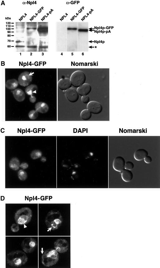
Npl4p-GFP localizes to ER/nuclear membranes. (A) Anti-Npl4 (left) and anti-GFP (right) Western blots of whole cell extracts derived from strains expressing endogenous Npl4p (lanes 1 and 4), a genomically tagged Npl4p-GFP (lanes 2 and 5), and a genomically tagged Npl4p-protein A(pA) (lanes 3 and 6). The anti-Npl4 blot shows the precise replacement of endogenous Npl4p with the two different C-terminally tagged proteins (compare lanes 2 and 3 to lane 1). The robust reactivity of anti-Npl4 with Npl4-pA (lane 3) is a result of antibody interactions with Npl4p epitopes as well as the IgG-binding pA tag. The rabbit anti-GFP blot recognizes both Npl4p-GFP and Npl4p-pA (lanes 5 and 6, respectively). The identity of the different proteins is indicated by arrows. The asterisk indicates an unidentified cross-reacting protein. (B) Npl4p-GFP localization as observed by fluorescence microscopy. The live cells were viewed at midlog phase. Localization is mostly nuclear (white arrowhead) and cytoplasmic, with an apparent concentration at perinuclear membranes. The white arrow indicates an example of Npl4p-GFP localization to peripheral membranes. A Nomarski image of the cells is shown to the right. (C) Npl4p-GFP colocalization with DAPI-stained nuclear DNA. Cells were fixed in formaldehyde, permeabilized with Triton X-100, and DAPI stained before fluorescence microscopy. The majority of Npl4p-GFP localizes to a region overlapping with and surrounding the nucleus as observed by DAPI staining. A Nomarski image of the cells is shown to the right. (D) Npl4p-GFP expression as viewed by fluorescence microscopy on a DeltaVision platform. Sequential 0.1-μm fluorescence images were taken through a diploid yeast strain expressing Npl4p-GFP from both NPL4 loci and subjected to five rounds of deconvolution. The images presented represent a central plane from deconvolved data. The white arrowhead indicates an example of Npl4p-GFP concentration at perinuclear membranes. White arrows indicate examples of Npl4p-GFP localization to peripheral membranes.

