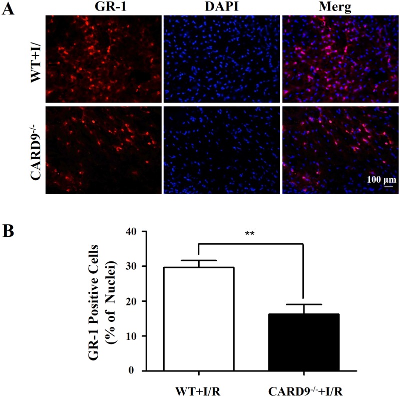
Fig 2. Immunofluorescence staining of neutrophils infiltrated in the heart tissue.
Following 45 min LAD occlusion and 24-h reperfusion, hearts from WT and CARD9-/- mice were cryo-sectioned (7 μm) and stained with antibodies against GR-1 for neutrophils (red) and DAPI for nuclei (blue). A, Representative heart sections showing GR-1, DAPI and merged staining images of neutrophils and nuclei; B, Analyses of the number of neutrophils as percentage of the total cell nuclei in the section. The number of infiltrated neutrophils in the WT mouse heart was significantly higher than that in the CARD9-/- mouse heart. Mean ± SEM, n = 3/group, **p < 0.01 vs. WT.

