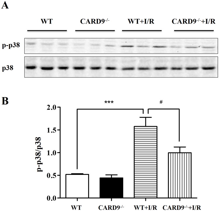
Fig 4. Western immunoblotting analyses of protein expressions in the heart.
Following reperfusion, heart tissue from the risk area was harvested and homogenized. Western immunoblotting analyses were performed to measure the expression levels of phospho-p38 MAPK and p38 MAPK from WT and CARD9-/- sham controls, WT+I/R, and CARD9-/-+I/R mice. A, Representative immunoblots of p-p38 MAPK and p38 MAPK; B, statistical analyses of the ratio of p-p38 MAPK/p38 MAPK. The phosphorylation level of p-38 MAPK was dramatically increased following I/R injury compared to sham controls. However, the ratio was significantly attenuated in CARD9-/-+I/R mouse heart compared to WT+I/R mouse heart. Mean ± SEM, n = 6/group, ***p < 0.001 vs. sham control; #p < 0.05 vs. WT.

