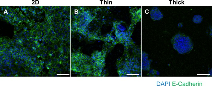
Fig 3. Immunofluorescent staining of E-Cadherin and DAPI to visualize syncytial fusion.
BeWo cells grown on (A) 2D, (B) thin Matrigel, or (C) thick Matrigel surfaces. Green fluorescence indicates E-Cadherin staining and blue fluorescence indicates DAPI staining for cell nuclei. Images were taken at 20x magnification and scale bar indicates 100 μm.

