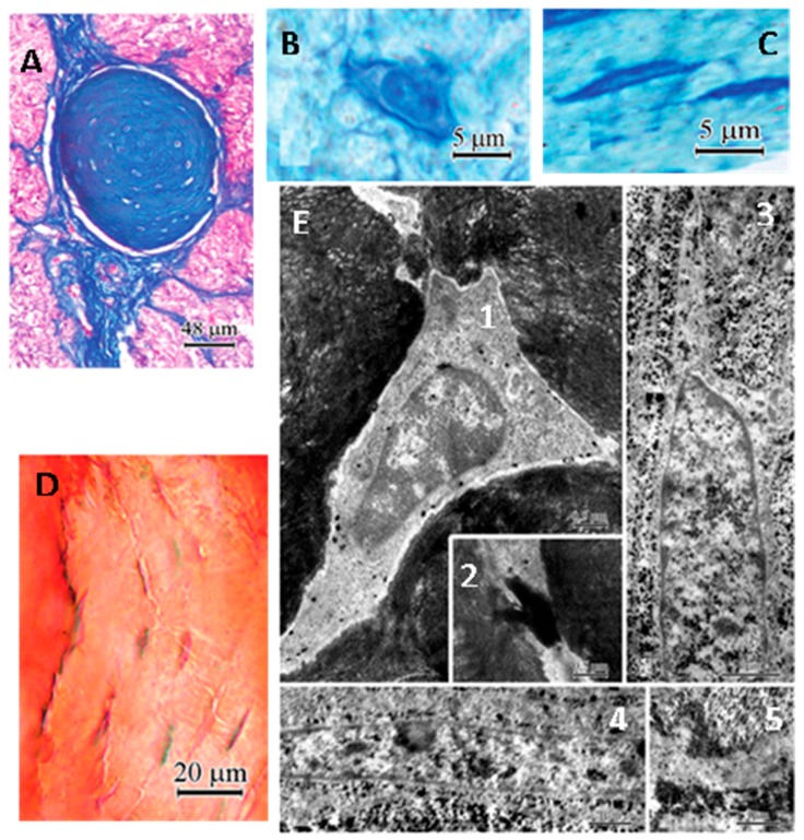
Figure 5.
Histology and ultrastructure of cells located in a concretion of the turkey pineal gland. (A) mature concretions identified with Mallory’s stain. Two types of cells are present in the concretion (semi-thin section stained with toluidine blue); (B) polygonal cells; (C) elongated cells; (D) The presence of calcium in the concretions was demonstrated with Alizarin red S; note the characteristic appearance of cells located in the concretion; (E) ultrastructure of cells located in the calcified area (fixation with the PPA method). 1: the osteocyte-like cell surrounded by mineralized collagen fibrils in the central part of the calcification area. Note the “halo” around the cell and large pyroantimonate precipitate located mainly outside the cell membrane, 2: the junction of processes of osteocyte-like cells, 3: a cell showing a fibrocyte-like appearance in the peripheral part of the calcification area. Note the extra-cellular matrix containing collagen and calcium deposits, 4: numerous calcium precipitates in the intercellular spaces in the peripheral part of the calcification area, 5: The cell process with scattered deposits in the middle part of the concretion. Note the adjacent extra-cellular matrix rich in collagen and pyroantimonate precipitates. Modified from Przybylska-Gornowicz et al. [159].

