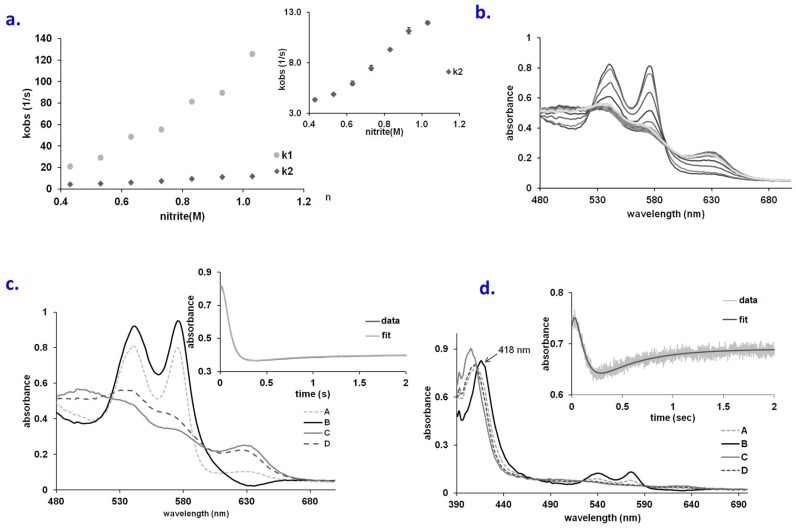
Figure 2.
(a) Plots of k1 and k2 vs. NO2− concentration for the reaction of oxyHb with NO2− at pH 7.4, aerobic; (b) UV-vis spectra collected upon mixing oxyHb (66.6 µM) and guanidine (1M) with NaNO2 (66 mM). Conditions: pH 7.4, PBS buffer, aerobic, over a range of 2 s; (c) Computed spectra for the species involved in the A → B → C → D reaction model (A—oxy Hb, B—Fe(II)-peroxynitrate, C-metHb, D—metnitriteHb). Conditions: 66.6 µM Hb, 1 M guanidine, 66 mM nitrite, pH 7.4, PBS buffer. Inset: fitting at 575 nm trace for the A → B → C → D kinetic model; (d) Computed spectra for the species involved in the A → B → C → D reaction model (A—oxy Hb, B—Fe(II)-peroxynitrate, C—metHb, D—met-nitriteHb). Conditions: 6.6 µM Hb, 33 mM nitrite, pH 7.4, PBS buffer, aerobic. Inset: fitting at 575 nm trace for the A → B → C → D kinetic model.

