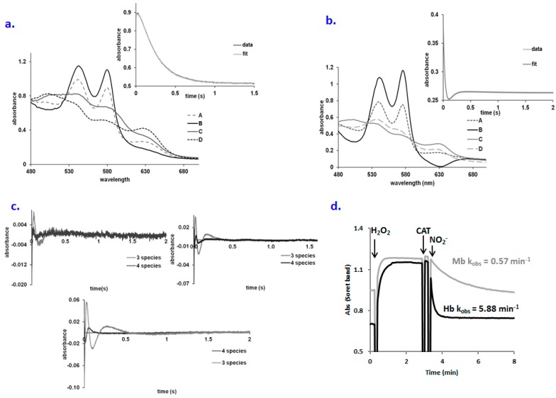
Figure 3.
(a) Computed spectra for the species involved in the A → B → C → D reaction model for oxyMb + nitrite (A—oxyMb, B—Fe(II)-peroxynitrate, C—metMb, D—met-nitriteMb). Conditions: 75 µM Mb, 0.5 M pH 7.4, PBS buffer, aerobic. Inset: fitting at 575 nm trace for the A → B → C → D kinetic model; (b) Computed spectra for the species involved in the A → B → C → D reaction model for oxyMb + nitrite (A—oxyMb, B—Fe(II)-peroxynitrate, C—metMb, D—met-nitriteMb). Conditions: 75 µM Mb, 0.5 M pH 7.4, PBS buffer, aerobic. Inset: fitting at 575 nm trace for the A → B → C → D kinetic model; (c) Residual plots of the A → B → C kinetic model vs. A → B → C → D kinetic model for the: left panel: oxy Mb + nitrite, right panel: oxyHb +nitrite, lower panel: oxyHb-guanidine + nitrite. Conditions cf. Materials and Methods; (d) Ferryl formation from the met form and its reduction with nitrite for both Mb and Hb (8 µM protein, 100 µM hydrogen peroxide, 0.3 µM catalase (CAT) for excess of peroxide removal, 0.3 mM nitrite, PBS buffer pH 7.4).

