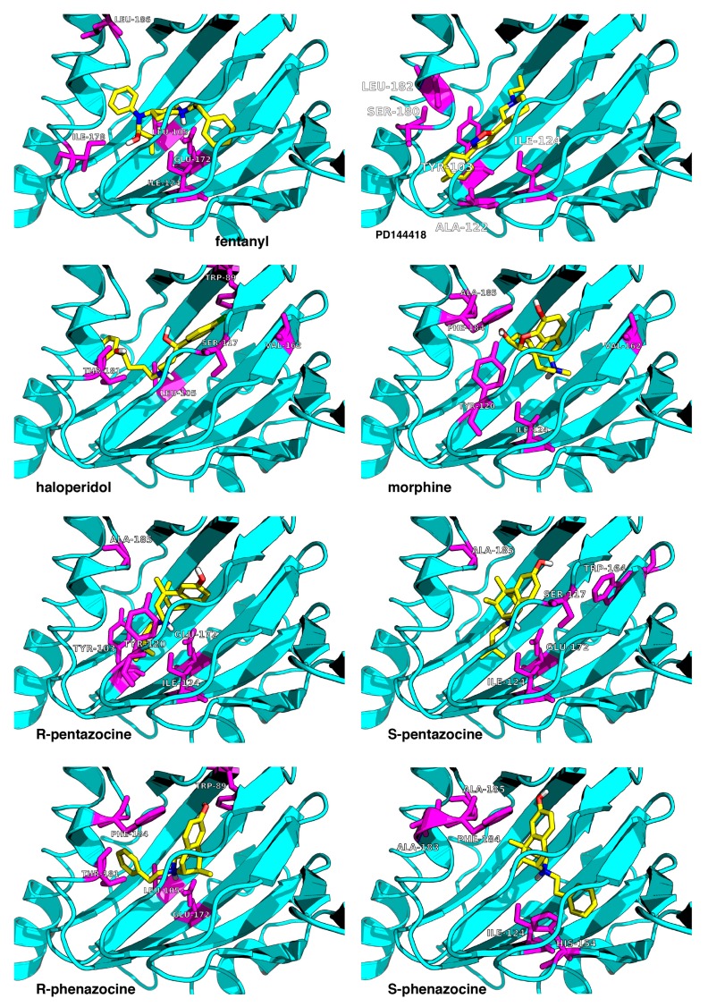
Figure 2.
Top-scored docking poses for the seven studied ligands (yellow) bound to the receptor (cyan). The eighth ligand is PD144418 (top right corner). The receptor’s residues most frequently contacted during the MD simulations are shown in magenta. The image is a zoomed view of the receptor binding site.

