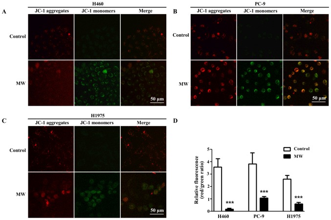
Figure 6.
Microwave (MW) hyperthermia treatment alters mitochondrial membrane potential (MMP). Cells were seeded onto 6-well plate at 2×10⁵/well and then exposed to a water bath or MW radiation at 43°C for 90 min. Following incubation for 6 h, MMP was examined by JC-1 staining with a confocal microscope. (A-C) Representative photographs from DCF fluorescence staining in the H460, PC-9 and H1975 cells, respectively. Scale bars, 50 µm. (D) Quantitative analyses of relative fluorescence are shown. ***P<0.001 compared to each control group.

