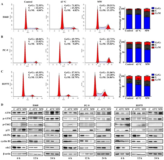
Figure 7.
Microwave (MW) hyperthermia induces G2/M arrest. Cells were seeded onto 6-well plate at 1×10⁶/well and then exposed to a water bath or MW at 43°C for 90 min. The cells were cultivated for 24 h, cell cycle distribution were analyzed by flow cytometry. (A-C) The cell cycle distributions following treatment of H460, PC-9 and H1975 cells. (D) Western blot analysis of ATM, p-ATM, p-Chk2, p21, cdc25c, cyclin B1 and cdc2 in NSCLCs cells. β-actin expression was included as an internal control. Values were the means of triplicate analyses.

