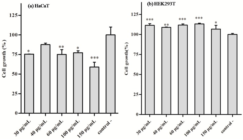
Figure 6.
The different concentrations of BF-30-loaded microspheres were used to determine the cytotoxicity. The cell growth of 4-arm-PEG-PLGA microspheres loaded with BF-30 using human HaCaT keratinocyte cells (a) and human HEK293T embryonic kidney cells (b). * p < 0.05, ** p < 0.01 and *** p < 0.001 versus microspheres without compound (negative control). Values are expressed as the mean ± SD of three independent experiments.

