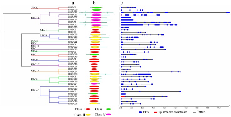
Figure 1.
Phylogenetic relationships, architecture of conserved protein motifs and gene structure in DlUBC genes from longan. (a) The neighbor-joining (NJ) tree on the left includes 40 DlUBC proteins from longan; (b) The architecture of conserved protein motifs of DlUBC proteins with the name of each corresponding protein is shown on the left. The position of the UBC domain is indicated in the panel. The different colors indicate the four E2 subtypes of the UBC domains; (c) Exon/intron structures of UBC genes from longan. Untranslated 5′ and 3′ regions, exon(s), and intron(s) are represented by red, blue boxes, and gray lines, respectively. The scale bar represents 1000 bp.

