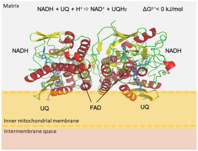
Figure 1.
Oxidoreduction reaction of NDH-2 enzymes. Cartoon representation of the model structure of the NDH-2 from L. infantum. The homodimer representation of LiNDH2 homology model generated using Phyre2 server is presented in a cartoon style. Orientation and structural arrangement of the monomer units were obtained using 3D alignment with the used template structures (Protein Data Bank IDs: 4g6g, 4g73 and 5jwa [9,10]). The homodimer consists of reciprocally oriented monomers and is located on the surface of the mitochondria intermembrane. C-termini are embedded into the membrane layer (inner mitochondrial membrane). In depicted reaction: NADH/NAD+ denote nicotinamide adenine dinucleotide protonated/deprotonated; UQ/UQH2 ubiquinone deprotonated/protonated and FAD flavin adenine dinucleotide.

