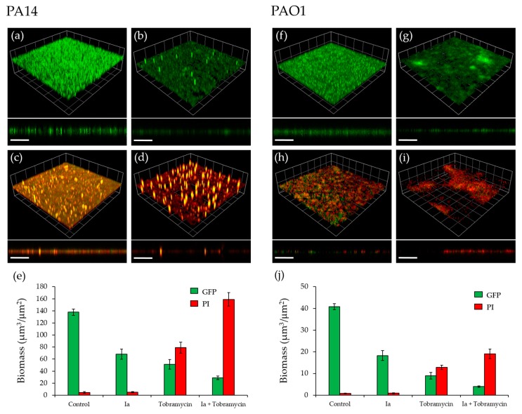
Figure 6.
Effect of Ia on P. aeruginosa biofilms. (a) Untreated GFP-labelled PA14 biofilm; (b) GFP- labelled PA14 biofilm grown with Ia at 8 µM; (c) GFP-labelled PA14 biofilm, treated with 100 µg/mL tobramycin for 4 h after 16 h of growth; (d) GFP-labelled PA14 biofilm grown with 8 µM Ia and treated with 100 µg/mL tobramycin for 4 h after 16 h of growth; (e) Biomass quantitation of PA14 biofilms; (f) Untreated GFP-labelled PAO1-L biofilm; (g) GFP-labelled PAO1-L biofilm grown with Ia at 34 µM; (h) GFP-labelled PAO1-L biofilm, treated with 100 µg/mL tobramycin for 4 h after 16 h of growth; (i) GFP-labelled PAO1-L biofilm grown with 34 µM Ia and treated with 100 µg/mL tobramycin for 4 h after 16 h of incubation; (j) Biomass quantitation of PAO1-L biofilms. Dead cells and extracellular DNA were stained with propidium iodide (PI). Three-dimensional (3D) sections and cross sections are shown. Scale bars represent 100 µm.

